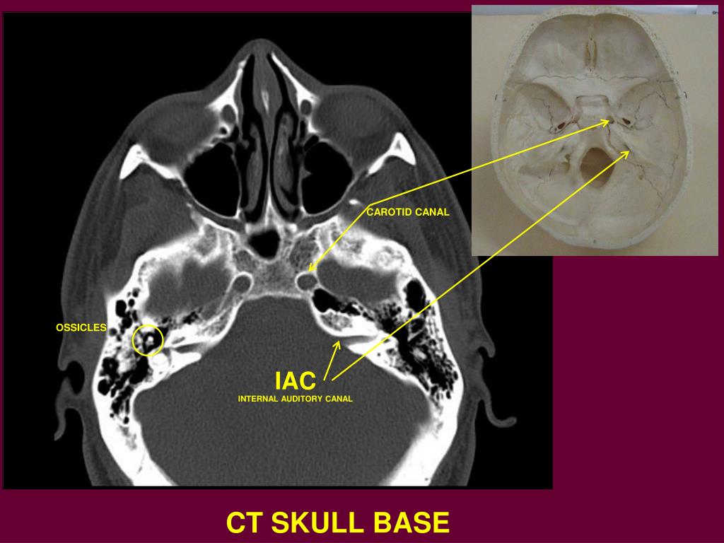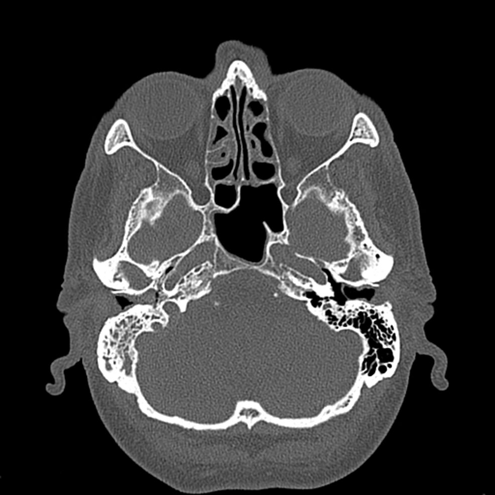
These measurements though approximated here, are critical to determination of constriction of these nerves from congenital conditions (Fatterpekar et al. The diameter of the 4 canals (approximately) making up the IAM are a shade under one millimeter with the exception of the canal of the auditory nerve which is larger (~2 mm). In examining a Stenver’s view of the IAM the nerve groupings can be placed into four quadrants (canals) following the location scheme mentioned earlier. This would show a cross section of the right IAM (see figure 1). This a lateral radiologic view across the head so the image aimed at the left side of the head would be of the right IAM. One of the best and most common ways to view the IAM and associated nerves in cross section is by a Stenver’s view. (Musiek and Baran, 2007 Fatterpekar et al.,1999). These can be divided into superior and inferior segments. In the posterior aspect of the IAM is the vestibular nerves. The auditory nerve is about 22 mm in length and as it courses through the IAM it twists clockwise slightly before entering the brainstem. The auditory nerve is positioned beneath the facial, hence it is anterior and inferiorly located in the IAM. The facial nerve proper carries mostly motor fibers and the nervous intermedius is composed of mostly sensory fibers. Within the IAM the facial can be observed to have two segments the facial nerve proper and the nervous intermedius. The facial nerve is located superior and anterior within the IAM. The position of these nerves is of import. These cranial nerves are longer than the IAM as they extend past the ends of the IAM. The facial, auditory and vestibular nerves course its length before exiting the temporal bone and projecting across the cerebellopontine angle into the lateral aspect of the brainstem at the ponto-medullary junction. In adult humans, the IAM is just under a centimeter (cm) in length and about 4 millimeters (mm) in diameter. Many audiologists either directly or indirectly assess these cranial nerves in their daily activities as clinicians.


These actual compose two of the cranial nerves – number 7 (facial) and 8 (auditory and vestibular).

This structure is germane to audiologists because it contains three nerves of interest to audiologists: 1- the auditory nerve, 2- the vestibular nerves, and 3- the facial nerve. The internal auditory meatus (IAM) is a canal in the temporal bone that extends from the bony cochlea medially to an opening in the posterior aspect of the petrous portion of the temporal bone.


 0 kommentar(er)
